
Internal Sinus Lift Through Six Year Follow-up
Part 6
The final Internal Sinus Lift image is of an unclouded healthy sinus with minimal to no dead space, buccal and palatal after five years. Graft material was Beta TCP.
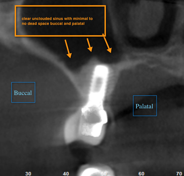
Mini study provided by Dr. Sheldon Lerner, DMD
Part 5
At five year follow up the patient has lost premolar, and desires an implant in #4. Upcoming part six will be a CT image exploring new site #5. It will give incredible views of what has happened to the sinus in the area of #3.
Mini study provided by Dr. Sheldon Lerner, DMD
Part 4
Image of crown on model in 2006. The next post will show five year post-op with CT follow up.
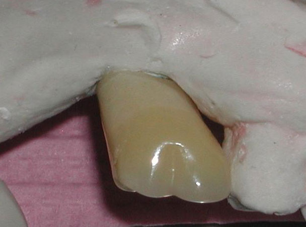
Mini study provided by Dr. Sheldon Lerner, DMD
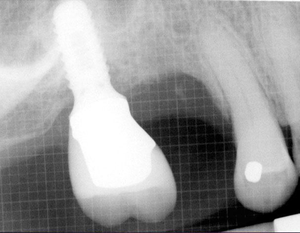
Part 3
Image is immediate post-op of internal sinus lift 2006 about 7mm lift on distal.
Mini study provided by Dr. Sheldon Lerner, DMD
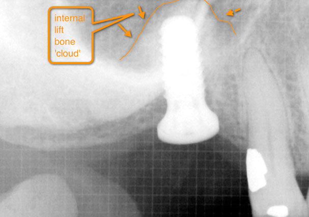
Part 2
Image is mid procedure, 2006. The initial bone height is 4-5mm. The 'cloud' above is the sinus lift.
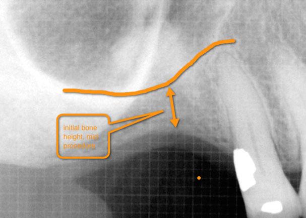
Mini study provided by Dr. Sheldon Lerner, DMD
Part 1
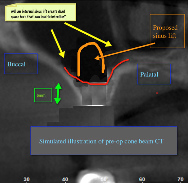
Part 1
Internal sinus lifts of 6mm or more are often criticized for possibly creating dead space on the buccal and lingual walls. Lateral lifts, which allow for direct visualization, are often chosen for the ability to directly lift the membrane and control dead space. This image is an illustration of the pre-op first molar site prior to implant placement.
Mini study provided by Dr. Sheldon Lerner, DMD